
1 – Nerve Conductions Studies (NCS)
2 – Electromyography (EMG)
3 – Electro-encephalography (EEG)
4 – Ambulatory EEG
5 – Video-telemetry EEG monitoring
6 – Visual evoked potentials (VEP)
7 – Brainstem Auditory Evoked Potentials (BAEP)
8 – Somatosensory Evoked Potentials (SSEP)
9 – Motor Evoked potentials (MEP)
10 – Intra-operative Neuro-physiologic Monitoring (IONM)
Nerve Conductions Studies (NCS)
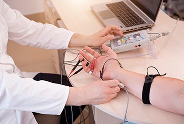
These are electrical tests which are used to test the function of peripheral nerves in the body. It measures function of sensory nerves (which transmit sensation from the peripheries to the brain) and these also test motor nerves (which transmit impulses from brain leading in muscle action). The tests provide useful information in diagnosis and prognosis of peripheral nerve disorders. The tests provide information on localisation, nature and severity of nerve disorders and injuries. The tests can sometimes provide information on prognosis of nerve disorders and injuries. During the test, surface recording electrodes are attached to skin or the over the muscles supplied by the particular nerve. They are either stick-on or Velcro-on pads. The hand held stimulating electrode is placed on skin over the area of the nerve to be tested. Brief electrical pulses are delivered by the stimulating electrode. The pulses can be slightly uncomfortable but are generally not too painful. The recording electrode picks up the impulses conducted through the nerves. Distance between the stimulating and the recording electrodes is measured by the recording physician or physiologist. Nerve conduction studies machine calculates time taken for the impulse to travel and conduction velocity and also measures size of the transmission response wave. The recording physician then analyses the data and formulate results. NCS is generally a safe procedure. You should inform neurophysiologist if you have a cardiac pacemaker or any electrical devices fitted. You can download patient information leaflet for further information.
Electromyography (EMG)
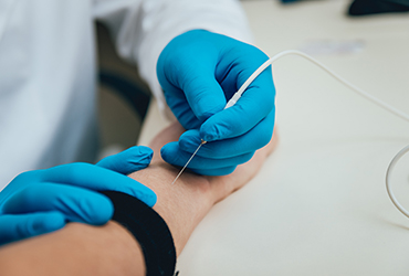
The tests provide information on function of muscle and the motor nerves supplying the muscles. The tests provide useful information on diagnosis and prognosis of nerve disorders, muscle disorders. During the test a fine needle electrode is inserted into a muscle. The needle records electrical signals from within the muscle. No electrical pulses are delivered. After the initial needle prick sensation, the test is generally painless and well tolerated. The recoding physician records electrical activity when the muscle is at rest. The recording physician then asks the patient to contract the muscle and records electrical activity while doing so. The recording physician then analyses the data and formulate results. EMG is generally a safe procedure. You should inform neurophysiologist if you regularly take blood thinning medications such as Asprin, Clopidogrel, Warfarin, Apixaban and like medications. You can download patient information leaflet for further information.
Electro-encephalography (EEG)
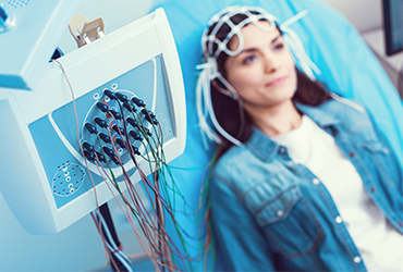
The test provides information about the function of brain. The test involves recording electrical activity generated by brain cells from the surface of brain. The tests provide useful information on diagnostic and prognostic information in certain conditions including seizure disorders, epilepsy, brain injuries, states of brain dysfunction and coma and other brain disorders. The test is carried out by qualified physiologists who attach wires with small metal disc electrodes at the ends to the scalp. The wires are then connected to hardware with an amplifier attached with a computer. No electric pulses are delivered and electrodes do not transmit any sensations. These just record your brain wave activity. The brain wave activity recorded shows up as wavy lines on an EEG recording. Once the electrodes and wires are attached, the recording is usually completed in 20 -60 minutes depending on requirements of the test. Physician neurophysiologist formulates and issues a report after analysing the results of the recording. The test is generally safe and painless and well tolerated. However, in order to increase the diagnostic yield of the test, certain exercises such as stimulation with strobe lights and breathing exercises are carried out in order to provoke epileptic changes in the recording. With these exercises, there remains a small risk of triggering a seizure during the test in patients with epilepsy, but appropriate medical supervision and care is provided if required
Ambulatory EEG
Ambulatory EEG test is similar to a standard EEG test in recording electrical brain wave activity but allows recording for longer periods of up to several days, typically 48-72 hours. The recording continues when the patient is at home and is asleep. This test increases the chances of catching an epileptic seizure or epilepsy related activity.
Video-telemetry EEG monitoring
This test is carried out in a similar way to standard EEG test but involves admitting the patient in a hospital setting. The test allows continuous and prolonged recording of electrical brain wave activity and simultaneous video recording, over a period of several days, typically 3-5 days. This test is used in complex and difficult to diagnose patients with suspected epilepsy and in epilepsy patients who need workup before considering epilepsy surgery. This is also a better test in differentiating epileptic seizures from non-epileptic seizure like episodes.
Visual Evoked Potentials (VEP)
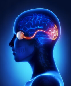
This is the most commonly used evoked potential test in clinical practice. The test involves record electrical activity transmitted and received in the brain in response to stimulation of eyes by visual stimuli. The test is used to diagnose disorders that can affect optic nerves which transmit visual signals from eyes to the brain. A trained healthcare professional attaches recording electrodes over the scalp. Eyes and the optic nerves are stimulated by asking patient to watch a checkerboard pattern on a screen for several minutes while electrical signals transmitted to the brain are recorded from the electrodes placed over the scalp. The test is generally safe, painless and well tolerated. Patient information leaflet is available for downloading for further information.
Brainstem Auditory Evoked Potentials (BAEP)
This test can diagnose hearing ability and can point to possible brainstem tumours or multiple sclerosis. A healthcare professional places electrodes on your scalp and earlobes and delivers auditory stimuli, such as clicking noises and tones, to one ear.
Somatosensory Evoked Potentials (SSEP)
This test can detect problems with the spinal cord that cause numbness of the arms and legs. For this test a healthcare professional attaches electrodes to your wrist, the back of your knee, or other locations. He or she will apply a mild electrical stimulus through the electrodes. Electrodes on your scalp then determine the amount of time it takes for the current to travel along the nerves to the brain.
Motor Evoked potentials (MEP)
For this test, a trained healthcare professional delivers electrical or magnetic stimuli to the brain. A trained healthcare professional attaches electrodes over the muscles and records electrical signals transmitted to muscles from the brain. Neurophysiology consultant physician interprets the data and formulates a report.
The tests are mainly used to assess the integrity of motor pathways that conduct signals from the brain to the muscles, through peripheral nerves and spinal cord and helps in the diagnosis of various neurologic conditions that can affect motor pathways. These tests are also used by trained neurophysiology technologists and physicians in the monitoring of motor pathways during complex brain and spinal surgeries.
Intra-operative Neuro-physiologic Monitoring (IONM)
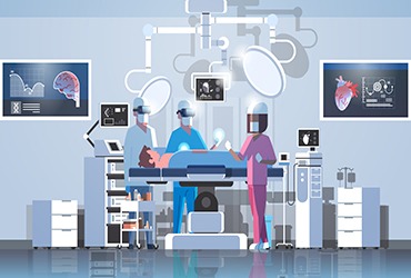
These tests are used during complex brain, brainstem and spinal surgeries, carried out by trained neurophysiology technologists and physicians. The tests provide useful information to the brain and spinal surgeons, about mapping of various brain and spinal cord regions and monitoring of various brain, brainstem and spinal cord pathways. The tests comprise various neurophysiologic modalities including motor evoked potential, somatosensory evoked potentials, brainstem auditory evoked potential, visual evoked potentials, electrocorticography and electromyography.